
Participants 426


The diagnosis generally requires the use of chest imaging in patients with compatible common CAP clinical presentations such as fever, dyspnea, cough, and sputum production.
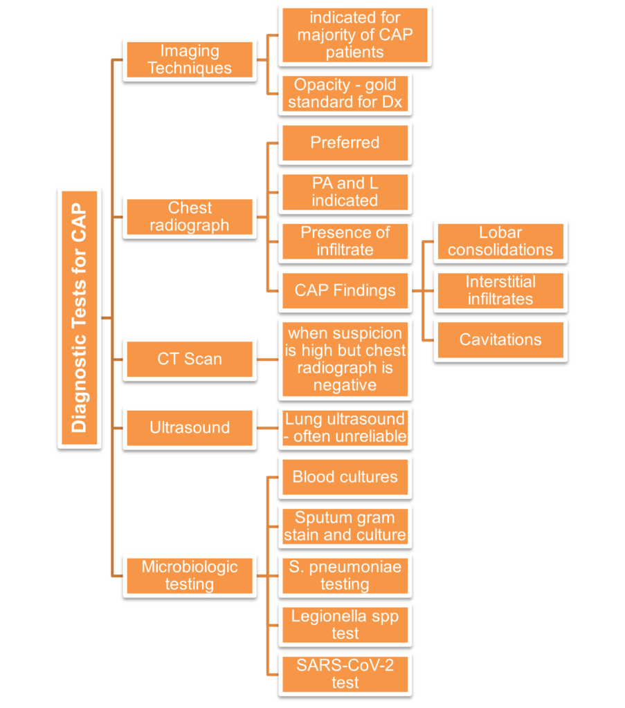
2. Chest radiograph – preferred main diagnostic method for CAP. Most patients would need posteroanterior and lateral chest radiographs. This is a necessity for hospitalized patients. Some radiographic findings consistent with CAP include:
· Lobar consolidations
· Interstitial infiltrates
· Cavitations
3. CT scan – done when clinical suspicion of CAP is high despite a negative chest radiograph as high resolution CT is more sensitive in terms of detection of pneumonia.
4. Ultrasound and other studies – lung ultrasound can also diagnose pneumonia particularly in unstable patients in the ED or ICU with difficulty in obtaining good-quality chest radiographs. However, this largely depends on the experience of the sonographer, therefore is not likely to be as reliable.
5. Microbiologic testing – aside from firm diagnosis with regards to presence of CAP pathogens, this helps with determining an empiric antibiotic therapy that will work efficiently for the patient. Obtain blood cultures, sputum gram stain and culture, urinary antigen testing for S. pneumoniae, test for Legionella spp, SARS-COV-2 testing.